

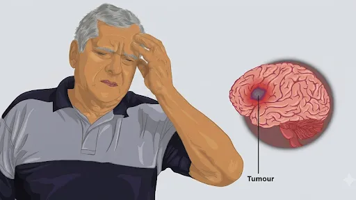
For decades, the battle against glioblastoma multiforme (GBM) has been one of the most disheartening fronts in oncology. As one of the most aggressive and lethal forms of brain cancer, its reputation is built on a terrifying trifecta: it spreads with predatory speed, resists the most potent treatments available, and is notoriously difficult to excise surgically. Even when surgeons successfully remove a visible tumor and follow up with a grueling regimen of radiation and chemotherapy, the victory is almost always temporary. Patients typically survive only six months to a year post-diagnosis. When the tumor returns—and it nearly always does—it is a more formidable foe: faster-growing, more resistant to drugs, and more adept at infiltrating the delicate architecture of the brain.
However, groundbreaking research from the Indian Institute of Technology Bombay (IIT Bombay), in collaboration with the Tata Memorial Centre’s ACTREC and the Indian Institute of Science (IISc), suggests that our failure to stop this relapse may stem from a fundamental flaw in how we study the disease. For over a century, cancer research has been conducted on rigid plastic Petri dishes. But the human brain is not rigid; it is one of the softest tissues in the body, possessing a consistency akin to jelly.
A series of studies led by Professor Abhijit Majumder of IIT Bombay’s Mechanobiology Laboratory reveals that this "physical mismatch" between the lab and the body is masking critical cancer-promoting genes and hiding potential drug targets. By recreating the "softness" of the brain, researchers have finally unmasked why glioblastoma is so resilient, offering a new paradigm for drug discovery.
I. Main Facts: The Mechanobiology of Cancer Relapse
The core of the discovery lies in the field of mechanobiology—the study of how physical forces and the mechanical properties of tissues affect cell behavior. The research team posits that the physical environment of a tumor is just as influential as its genetic mutations.
The "Invisible" Aggression
The researchers discovered that when brain cancer cells are grown on standard laboratory plastic, the distinction between "parent" cells (the original tumor) and "recurrent" cells (those that survive treatment and cause relapse) vanishes. On plastic, both populations look and act the same. However, when placed on soft hydrogels that mimic the actual stiffness of the human brain, the recurrent cells undergo a dramatic transformation. They become elongated, highly mobile, and significantly more invasive.
The Discovery of PLEKHA7
By aligning the laboratory environment with the physical reality of the brain, the team identified a specific protein, PLEKHA7, which is significantly elevated in recurrent glioblastoma. Crucially, this protein only becomes visible as a target when cells are grown on brain-mimicking surfaces. In clinical biopsies of patients with relapsed GBM, PLEKHA7 levels were found to be high, confirming that the soft-surface model accurately reflects human pathology.
The Role of NEAT1 and lncRNAs
The research further identified NEAT1, a long non-coding RNA (lncRNA), as a "mechanosensor." This molecule translates the physical cues of tissue softness into biological signals that drive tumor growth and movement. NEAT1 levels were found to be three to four times higher in cells grown on soft surfaces compared to those on plastic, and even higher in 3D "tumoroid" models.
II. Chronology of Discovery: From Plastic to Physiology
The journey toward these findings involved several years of interdisciplinary collaboration, moving from simple cell cultures to complex 3D models.
Step 1: Recreating the Relapse (The ACTREC Collaboration)
The research began with a collaboration between Prof. Shilpee Dutt (formerly at ACTREC, Tata Memorial Centre, now at JNU) and Prof. Majumder’s team. To understand relapse, they had to create it in the lab. They exposed brain cancer cells to radiation, killing 90% of the population. The 10% that survived—the "recurrent" cells—were allowed to regrow.
When these survivors were implanted into mice, they were far more aggressive than the original cells. But when grown on standard lab plastic, they appeared identical to the original cells. This discrepancy led the team to suspect that the rigid plastic was "dulling" the cells’ true behavior.
Step 2: The Soft-Gel Breakthrough
To test this theory, the team engineered polyacrylamide hydrogels with a "Young’s modulus" (a measure of stiffness) matching that of brain tissue. As soon as the recurrent cells were placed on these soft gels, their aggressive nature—hidden for years by plastic dishes—became visible. This work was published in the journal Matrix Biology, highlighting that the physical environment, not just genetics, dictates how a tumor behaves after radiation.
Step 3: Identifying the Genetic Sensors (The IISc Collaboration)
Once the team established that stiffness mattered, they sought the "messenger" within the cell. Working with Prof. Mohit Kumar Jolly at IISc Bengaluru, Dr. Arpita Ghosh (the lead author of the follow-up studies) focused on NEAT1. They discovered that this RNA molecule was hyper-responsive to the softness of the environment. This phase of the research, presented in bioRxiv, proved that cancer cells "feel" their environment through specific molecular pathways.
Step 4: Moving into the Third Dimension
Finally, the researchers addressed the limitation of 2D surfaces. Real tumors are 3D "balls" of cells. In a study published in Macromolecular Bioscience, the team grew glioblastoma "tumoroids." They found that NEAT1 levels surged even further in 3D environments, suggesting that the architecture of the tumor itself is a driver of malignancy.
III. Supporting Data: Quantifying the Impact of Environment
The research provides compelling data points that explain why previous drug trials may have failed.
- Gene Expression Disparities: In the comparison between plastic and soft gels, PLEKHA7 was entirely absent or negligible on plastic but highly expressed on soft surfaces. When researchers blocked PLEKHA7 in the soft-gel models, the survival and spread of the cancer cells were significantly reduced.
- NEAT1 Amplification: On brain-mimicking gels, NEAT1 levels increased by 300% to 400% compared to plastic. In 3D tumoroid models, this increase was 3.5 times higher than even the 2D soft gels.
- Morphological Changes: Under the microscope, tumoroids with active NEAT1 displayed "jagged, protruding edges," a classic sign of invasive cells preparing to break away and migrate. When NEAT1 was suppressed using RNA interference, the tumoroids became "smooth and compact," indicating a loss of invasive potential.
- Cross-Cancer Validation: The team tested other cell lines (breast, lung, cervical, and liver). They found that while NEAT1 and a related RNA, MALAT1, increased in 3D models for breast and lung cancer, the response varied in liver and cervical cancers. This suggests that every cancer has a "mechanical signature" unique to the organ it inhabits.
IV. Official Responses: Insights from the Lead Researchers
The researchers emphasize that these findings are a wake-up call for the pharmaceutical industry and the broader scientific community.
Prof. Abhijit Majumder (IIT Bombay):
"We suspected that the problem lay in the experimental model itself. While the brain is one of the softest tissues in the body, cancer cells are routinely studied on rigid plastic surfaces. The rigid surface didn’t just dull the cells’ aggressive behaviour; it erased the very distinction between parent and recurrent cells that defines clinical relapse."
Prof. Shilpee Dutt (JNU/ACTREC):
"A real, functionally validated drug target [PLEKHA7] would have simply been missed using the conventional culture method most labs still rely on. If someone has a cancer drug target, they should now include soft-gels and tumoroids as an initial experiment."
Dr. Arpita Ghosh (Lead Researcher):
"The ability of cancer cells to self-renew, to spread through tissue, and to adopt a more mobile, shape-shifting form all went down when we suppressed NEAT1. This told us NEAT1 is a mechanosensitive molecule capable of translating physical cues into changes in tumour behaviour."
V. Implications: Why 90% of Cancer Drugs Fail
The implications of this body of work extend far beyond the laboratory. Currently, approximately nine out of ten cancer drug candidates fail in human clinical trials, despite showing immense promise in early lab testing. This staggering failure rate carries a massive human and financial cost.
1. Redefining Drug Screening
The IIT Bombay research suggests a primary reason for this failure: the "initial promise" of these drugs is often measured on plastic dishes that do not reflect the biological reality of the human body. If a drug target like PLEKHA7 is invisible on plastic, how many other potential cures have been discarded? Conversely, how many drugs that work on plastic fail in humans because the target isn’t actually relevant in the soft environment of the brain?
2. Personalized Mechanobiology
The discovery that different cancers (breast vs. liver) respond differently to 3D structures and stiffness suggests that oncology must move toward "organ-specific" testing environments. Testing a brain cancer drug on the same platform used for a bone cancer drug is increasingly seen as scientifically unsound.
3. A New Target for Therapy
The identification of NEAT1 and PLEKHA7 as mechanosensitive drivers of relapse opens a new door for therapeutic intervention. By targeting the molecules that allow cancer cells to "sense" and adapt to the brain’s soft environment, clinicians may finally be able to prevent the aggressive spread that characterizes glioblastoma relapse.
4. The Future of Lab Standards
The researchers are calling for a shift in global laboratory standards. While plastic dishes are "convenient, reproducible, and standardized," they are biologically "silent." The team argues that incorporating soft-gel substrates and 3D tumoroid models into the early stages of drug development is no longer a luxury—it is a necessity for clinical success.
By bridging the gap between physics and biology, the teams at IIT Bombay, ACTREC, and IISc have provided a roadmap that could finally turn the tide against glioblastoma, ensuring that the next generation of cancer treatments is built on a foundation that matches the reality of the human body.
