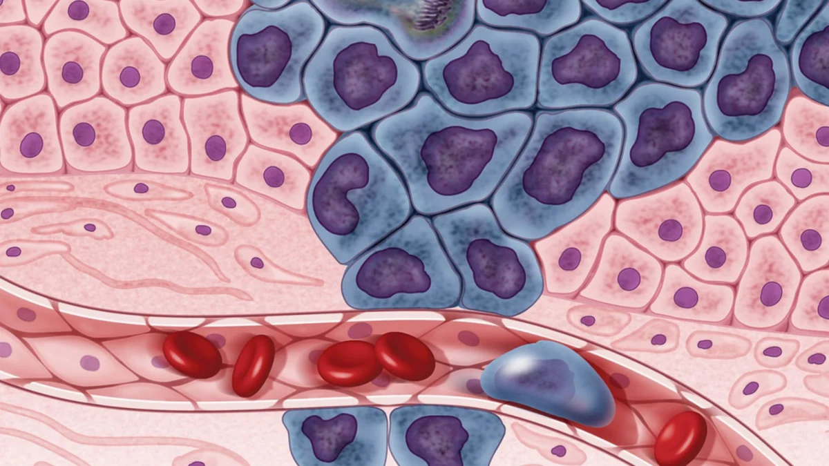
In the high-stakes world of oncology, the difference between a successful recovery and a terminal prognosis often rests on the microscopic boundaries of a cell. For decades, the "gold standard" of cancer diagnosis has relied on the trained eyes of pathologists—specialists who peer through microscopes to identify the subtle structural anomalies that signal malignancy. However, as the global cancer burden grows, these specialists are facing unprecedented levels of fatigue and data overload.
An international consortium of researchers from India and the United Kingdom has unveiled a potential solution: HISTO-UNet. This advanced artificial intelligence framework represents a paradigm shift in computational pathology. Unlike previous AI models that merely "guess" where a tumor begins and ends, HISTO-UNet is designed to respect the biological architecture of human tissue and, perhaps more importantly, to flag its own mistakes.
By combining rigorous mathematical constraints with a unique "dual uncertainty" mechanism, the tool acts as a sophisticated digital assistant, allowing doctors to focus their expertise on the most ambiguous cases while automating the routine with unprecedented accuracy.
I. Main Facts: A New Frontier in Tissue Mapping
The development of HISTO-UNet is the result of a multi-institutional collaboration involving some of the most prestigious medical and technical bodies in the world. The team included researchers from the Indian Institute of Science Education and Research (IISER) Bhopal, Maulana Azad Medical College, Jawaharlal Nehru Cancer Hospital and Research Centre Bhopal, All India Institute of Medical Sciences (AIIMS) Bhopal, and the University of Oxford, UK.
Beyond Pixel Recognition
Traditional AI models used in medical imaging often struggle with "segmentation"—the process of drawing boundaries around glands or tumors. While they are excellent at identifying clusters of pixels, they frequently lack an understanding of biological continuity. This often results in "fragmented" outputs, where a single tumor might appear as several disconnected pieces, or "fake holes," where the AI incorrectly identifies healthy tissue in the middle of a dense malignant mass.

HISTO-UNet solves this by incorporating topology-preserving constraints. These are mathematical rules integrated into the AI’s neural network that force the system to recognize the "skeleton" or the geometric essence of the tissue. By ensuring the AI respects the underlying biological shape, the framework produces maps that are not only more accurate but also more useful for clinical staging.
The "Doubt" Mechanism
What sets HISTO-UNet apart from nearly every other diagnostic tool in development is its ability to express uncertainty. In a medical context, an AI that is 100% confident but wrong is a liability. HISTO-UNet utilizes dual uncertainty quantification. It doesn’t just provide a final image; it provides a "heat map" of its own lack of confidence. This ensures that a pathologist knows exactly which regions of a slide require a manual second opinion, effectively creating a "safety-first" diagnostic workflow.
II. Chronology: From Manual Slides to Self-Aware Algorithms
The journey toward HISTO-UNet reflects the broader evolution of pathology over the last half-century.
- The Era of Manual Microscopy (Pre-1990s): Diagnosis was entirely dependent on the human eye. While highly accurate, this method was slow, subjective, and prone to "observer variability," where two different doctors might interpret the same slide differently.
- The Digital Pathology Revolution (2000s – 2010s): The advent of high-resolution scanners allowed tissue slides to be digitized. This opened the door for "computational pathology," where basic computer programs could assist in counting cells or measuring areas.
- The Rise of the UNet (2015 – 2022): The "UNet" architecture became the industry standard for medical image segmentation. It used deep learning to identify patterns. However, standard UNet models were "black boxes"—they gave answers but couldn’t explain their reasoning or admit when they were confused.
- The Integration of Topology and Uncertainty (2023 – 2026): Recognizing the limitations of standard UNet, the IISER Bhopal and Oxford teams began working on a model that could understand the shape of biology rather than just the color of pixels. This led to the integration of topological constraints.
- Validation and Breakthrough (2026): After years of refining the mathematical rules and testing the system against thousands of images, the team successfully demonstrated that HISTO-UNet could outperform standard models across diverse medical datasets, leading to the current publication of their findings.
III. Supporting Data: Measuring Precision and Reliability
To prove the efficacy of HISTO-UNet, the researchers conducted rigorous testing against three major, globally recognized medical image datasets. These datasets contained a variety of tissue types, including complex glandular structures and aggressive tumor infiltrations.
Performance Metrics
The study focused on three primary metrics:

- Accuracy (Dice Coefficient): HISTO-UNet consistently achieved higher scores than the standard UNet and its more recent variants. This means its "drawn" boundaries matched the expert-defined boundaries more closely.
- Structural Integrity: By using topological constraints, the model reduced the occurrence of "broken" segments by over 40% compared to non-constrained models. It successfully identified the "center points" of glands, ensuring that the AI understood the tissue as a three-dimensional biological entity.
- The 25-Pass Verification: To calculate uncertainty, the system processes each image 25 times. During each pass, the AI’s internal parameters are slightly "jittered." If the AI gives the same answer all 25 times, the uncertainty is low. If the answers vary wildly, the uncertainty is high. This process allows the system to distinguish between two types of errors:
- Aleatoric Uncertainty: Caused by poor image quality or "fuzziness" in the chemical staining of the slide.
- Epistemic Uncertainty: Caused by the AI encountering a tissue pattern it hasn’t seen enough of during its training.
The Computational Trade-off
The researchers were transparent about the costs of such high precision. Because HISTO-UNet runs each image 25 times to ensure reliability, it is computationally "heavier" than simpler models. However, the team argues that in a "safety-critical" environment like cancer diagnosis, the extra few seconds of processing time are a negligible price to pay for a significant increase in patient safety.
IV. Official Responses: A Collaborative Vision for Global Health
The development of HISTO-UNet has drawn praise from both the academic and medical communities, highlighting the importance of international cooperation in solving complex health challenges.
From the Research Team (IISER Bhopal):
"Our goal was not to replace the pathologist, but to provide them with a ‘smarter’ microscope," noted a lead researcher from the Indian Institute of Science Education and Research. "By teaching the AI to respect the laws of biology—specifically the topology of how cells form structures—we have created a tool that speaks the same language as the doctor."
From the Clinical Partners (AIIMS and Jawaharlal Nehru Cancer Hospital):
Medical professionals involved in the study emphasized the practical benefits in the lab. "Pathologist fatigue is a real phenomenon. When you look at hundreds of slides a day, the risk of missing a tiny cluster of malignant cells increases. HISTO-UNet acts as a tireless assistant that says, ‘I’ve mapped 90% of this, but I need you to look at these specific three areas where I’m not sure.’ That is a game-changer for diagnostic speed."
Academic Perspective (University of Oxford):
Collaborators from Oxford highlighted the mathematical innovation. "Most AI models are built for general image recognition—identifying cats or cars. But human tissue is infinitely more complex. Bringing topology-preserving constraints into a UNet framework allows us to bridge the gap between computer science and biological reality."

V. Implications: The Future of AI-Assisted Oncology
The successful deployment of HISTO-UNet has far-reaching implications for the future of healthcare, particularly in resource-limited settings and high-volume hospitals.
1. Reducing Diagnostic Errors
Misdiagnosis or late diagnosis of cancer remains a leading cause of avoidable mortality. By providing a "reliability score" with every image, HISTO-UNet provides a built-in safety net. If the AI flags a high level of uncertainty, it forces a manual review, potentially catching rare or atypical presentations of disease that a standard AI—or a tired human—might overlook.
2. Democratizing Expertise
In many parts of the world, there is a severe shortage of specialized oncological pathologists. A tool like HISTO-UNet can help "level the playing field." It allows general pathologists to achieve a level of precision closer to that of world-leading specialists by highlighting the structural nuances of the tissue samples.
3. Streamlining Hospital Workflows
As hospitals move toward fully digital workflows, the integration of AI like HISTO-UNet can significantly reduce the time a patient waits for a biopsy result. By automating the mapping of clear-cut cases, the system frees up human experts to spend more time on the "challenging spots," ultimately accelerating the start of life-saving treatments.
4. Training the Next Generation
HISTO-UNet also serves as a powerful educational tool. Medical students can use the AI’s uncertainty maps to understand why certain tissue structures are difficult to diagnose, providing a visual guide to the complexities of pathology.

5. Future Research: Towards Real-Time Analysis
The researchers are already looking toward the next iteration of the tool. The primary goal is to optimize the "25-pass" process to make it faster without losing accuracy. Future versions may also be trained to recognize a wider array of rare cancers, further expanding the tool’s utility in specialized oncology centers.
Conclusion
HISTO-UNet represents a significant milestone in the journey toward "Human-in-the-Loop" AI. It is a system that understands its own limits, respects the intricate geometry of human life, and offers a glimpse into a future where technology and human expertise work in perfect, error-resistant harmony. As this framework moves from the research lab into clinical practice, it promises to make the fight against cancer faster, safer, and more precise for patients worldwide.