
KANPUR, INDIA — In a landmark development for regenerative medicine, researchers from the Indian Institute of Technology (IIT) Kanpur and IIT Delhi have unveiled a breakthrough that could fundamentally alter the trajectory of joint health for millions. By combining the ancient resilience of silk with cutting-edge 3D-bioprinting technology, the team has successfully demonstrated a method to regrow functional knee cartilage in living organisms—a feat that has long eluded the medical community.
The study, recently published and conducted at the intersection of bioengineering and molecular biology, offers a glimmer of hope for the more than 500 million individuals worldwide suffering from osteoarthritis (OA). As the global population ages, the demand for non-invasive, permanent solutions to joint degradation has reached a fever pitch. This new research suggests that the answer may not lie in metal and plastic replacements, but in a sophisticated biological scaffold that "tricks" the body into healing itself.
The Main Facts: A Paradigm Shift in Cartilage Regeneration
Osteoarthritis is characterized by the progressive breakdown of hyaline cartilage—the smooth, white tissue that covers the ends of bones where they come together to form joints. Unlike skin or bone, cartilage is avascular, meaning it lacks a direct blood supply, which makes its natural capacity for self-repair almost non-existent. Once it wears away, the resulting bone-on-bone friction causes debilitating pain and loss of mobility.
The core of the IIT team’s innovation lies in a specialized 3D-printed implant. Key highlights of the discovery include:
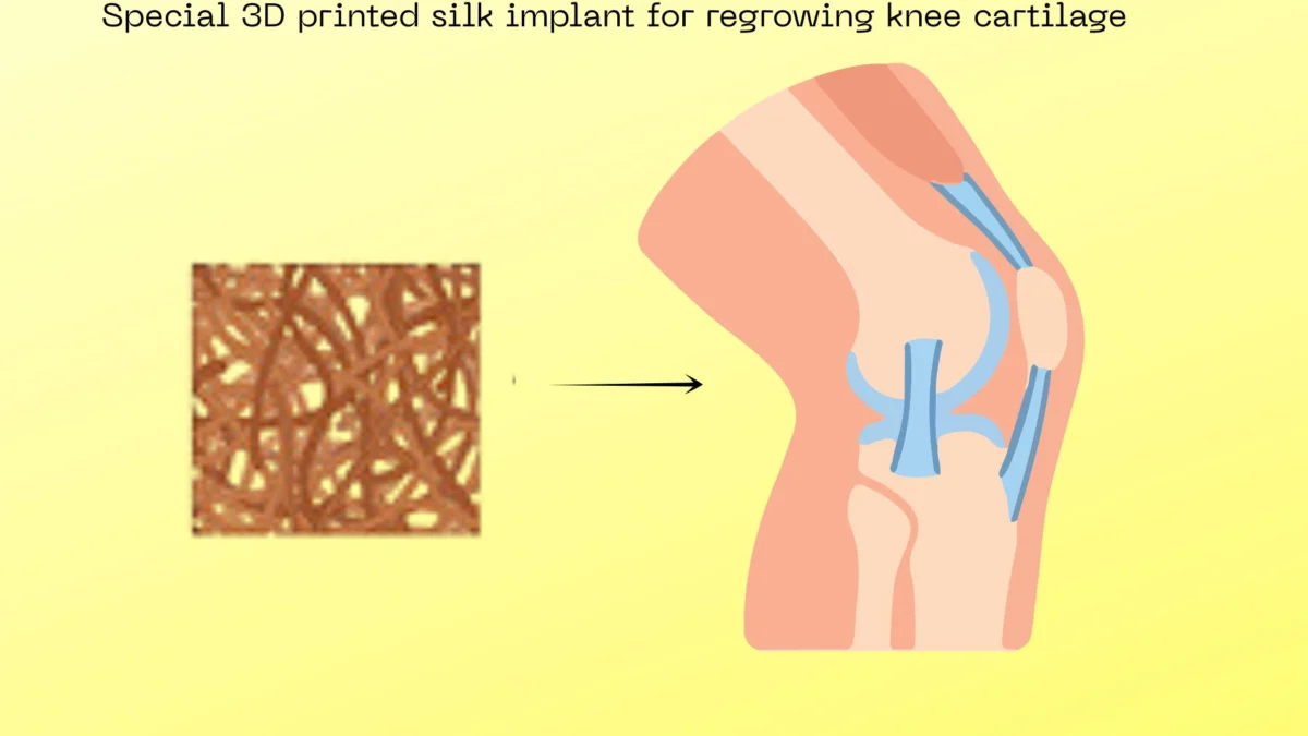
- Novel Bio-ink: A hybrid material composed of silk fibroin (derived from silkworms) and gelatin, providing both structural integrity and biological compatibility.
- Targeted Drug Delivery: The implant is chemically bonded with a specific molecule, LDN-193189, which inhibits harmful biological pathways that turn soft tissue into bone.
- Dual-Action Mechanism: While the drug prevents "bad" growth (hypertrophy), the silk itself promotes "good" growth (hyaline cartilage) by activating the Wnt signaling pathway in stem cells.
- Successful Animal Trials: In rat models with severe osteoarthritis, the implants facilitated the growth of healthy, functional cartilage within four months, whereas control groups showed continued joint degradation.
Chronology of the Breakthrough: From Lab to Living Tissue
The journey to this discovery began with a deep dive into the molecular "miscommunications" that occur within an osteoarthritic joint.

Phase 1: Identifying the BMP Pathway
The researchers first identified that in an osteoarthritic environment, the Bone Morphogenetic Protein (BMP) pathway becomes hyperactive. Under normal conditions, BMP helps build bone, but in a joint meant for flexible cartilage, this hyperactivity is disastrous. It causes chondrocytes (cartilage cells) to undergo hypertrophy, essentially turning flexible joint tissue into rigid, painful bone spurs.
Phase 2: Engineering the Scaffold
Recognizing that a simple physical plug would not suffice, the team turned to 3D printing. They developed a "bio-ink" using silk fibroin. Silk was chosen for its extraordinary mechanical strength and its ability to be resorbed by the body over time. By mixing it with gelatin, they created a substance that could be printed into precise, porous geometries designed to fit a patient’s specific injury.
Phase 3: The Chemical Marriage
The most critical step was the covalent bonding of the drug LDN-193189 to the silk matrix. Unlike previous attempts where drugs were simply "soaked" into an implant and washed away by joint fluid within hours, this chemical bond ensured a sustained, slow release. This "slow-drip" effect is essential for protecting the stem cells that migrate into the area over several weeks.
Phase 4: The Rat Model Study
To test the efficacy, the team induced severe osteoarthritis in rats. They then surgically cleared the damaged areas and inserted the 3D-printed constructs. Over a 120-day period, the researchers monitored the rats using high-resolution imaging and biochemical analysis to track the transformation of the implant into living tissue.
Supporting Data: Evidence of Regeneration
The evidence gathered during the four-month observation period provided a stark contrast between the experimental and control groups.

1. Stem Cell Migration and Survival:
Using chemical markers, the researchers tracked the movement of mesenchymal stem cells (MSCs) from the rats’ bone marrow into the silk implant. In the drug-laced scaffolds, these cells not only survived the "toxic" inflammatory environment of the osteoarthritic joint but also began to differentiate into healthy chondrocytes.
2. Micro-CT and Histological Analysis:
Micro-Computed Tomography (Micro-CT) scans revealed that the rats treated with the silk-drug implant had smooth, continuous joint surfaces. Histological staining (microscopic examination of tissue) confirmed the presence of Type II collagen—the hallmark of high-quality hyaline cartilage.
3. Comparison with Control Groups:
- Untreated Group: Showed significant bone-on-bone rubbing, increased inflammation, and the formation of osteophytes (bone spurs).
- Silk-Only Group (No Drug): While the silk provided some structural support, the lack of BMP inhibition meant the new tissue often turned into "fibrocartilage" (scar-like tissue) or underwent hypertrophy, eventually hardening into bone.
- Drug-Laced Group: This group was the only one to show the regeneration of true hyaline cartilage, mirroring the original tissue of a healthy knee.
Official Responses and Research Context
The research community has greeted these findings with cautious optimism. Spokespersons from IIT Kanpur emphasized that the multidisciplinary nature of the project—combining polymer science, pharmacology, and surgical expertise—was the key to its success.
"The challenge in cartilage engineering has never been just about making a scaffold," noted a senior researcher involved in the study. "It’s about managing the environment. By binding the inhibitor directly to the silk, we created a ‘safe zone’ for the body’s own stem cells to do their work. We aren’t just replacing the joint; we are directing a biological repair mission."

Outside experts in the field of regenerative medicine have pointed out that this study addresses one of the biggest hurdles in the field: the "washout" effect. "In the past, we’ve had great drugs that could stop osteoarthritis, but we couldn’t keep them in the joint long enough to work," says Dr. Aris Thorne, a specialist in orthopedic bioengineering (independent of the study). "This covalent bonding to a 3D-printed matrix is a sophisticated solution to a very old problem."
Implications: The Future of Orthopedic Care
The potential implications of this technology are vast, ranging from individual patient outcomes to global healthcare economics.
Moving Beyond Total Knee Replacement (TKR)
Currently, the "gold standard" for severe osteoarthritis is Total Knee Replacement. While effective, TKR is a major surgery with a long recovery time, and the prosthetic joints have a finite lifespan (usually 15–20 years). For younger patients, this often means facing a second, more difficult "revision" surgery later in life. The IIT Kanpur/Delhi implant offers a "biological fix" that could potentially last a lifetime, as the regenerated tissue is the patient’s own.
Economic Impact
The cost of joint replacement surgeries and the subsequent physical therapy puts a massive strain on healthcare systems. A 3D-printed solution, once scaled, could be significantly more cost-effective. Because the silk can be sourced from the textile industry and 3D printing allows for customized, on-demand manufacturing, the barrier to entry for this treatment could be much lower than traditional prosthetics.
The Road to Human Trials
Despite the success in rats, the researchers are quick to manage expectations regarding the timeline for human use. The next phase of research involves:
- Large Animal Trials: Testing the implants in dogs or pigs. These animals are "load-bearing," meaning their joints face stresses much closer to those of a human.
- Aging Studies: Osteoarthritis is primarily a disease of the elderly. The team needs to ensure that the older stem cells found in aging populations are as responsive to the silk-drug scaffold as the cells in their younger rat models.
- Immune Integration: Further study is required to see how the human immune system interacts with the silk fibroin over long periods.
Conclusion
The work coming out of IIT Kanpur and IIT Delhi represents a landmark achievement in the quest to cure osteoarthritis. By leveraging the mechanical properties of silk and the precision of 3D printing, and by solving the problem of drug delivery through covalent bonding, these researchers have provided a blueprint for the future of regenerative medicine.
If these results hold true in larger animals and eventually humans, the "silent epidemic" of joint pain may finally meet its match. Instead of a future of metallic implants and limited mobility, the next generation of aging adults may look forward to a world where their own bodies, guided by silk and science, can heal the very joints that carry them through life.
