

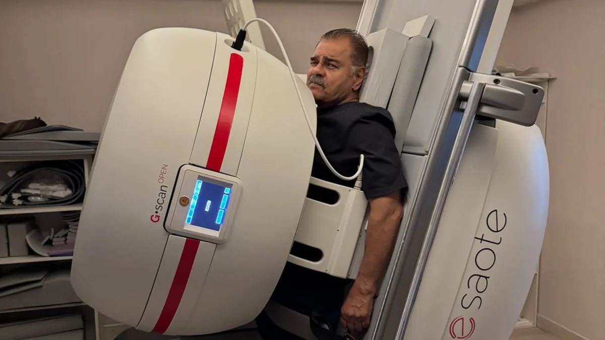
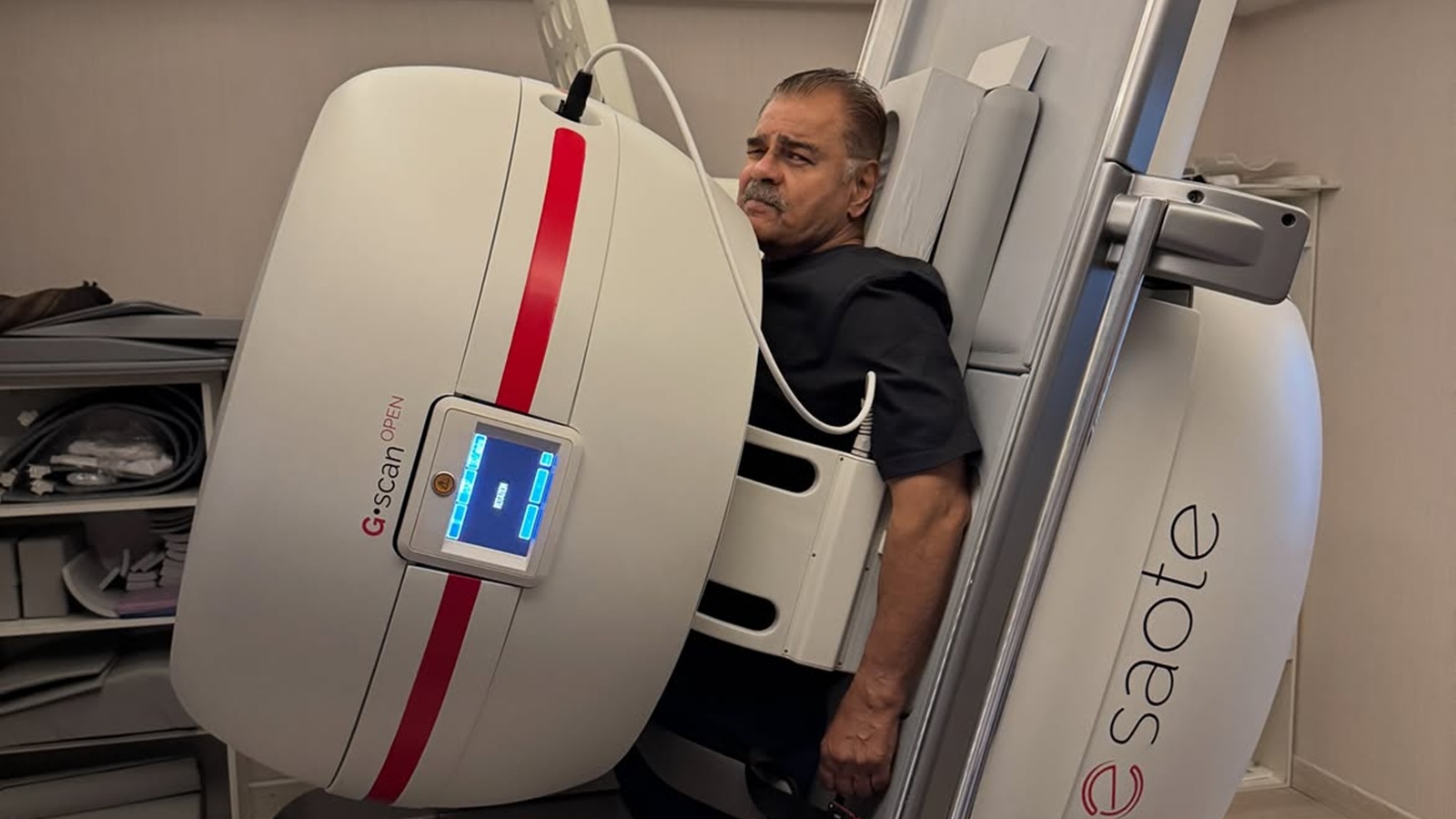
New Delhi, September 10, 2026 – Renowned for his impactful performances and distinctive screen presence, 76-year-old actor Sharat Saxena has recently brought a lesser-known but significant medical advancement into the public spotlight. Saxena shared on his Instagram that he underwent a "standing MRI before lumbar spine surgery" at Bombay Hospital. This disclosure, accompanied by a wave of well wishes from industry colleagues, underscores the evolving landscape of medical imaging, particularly for spinal conditions. His expressed hope to "walk properly again and be able to work" resonates deeply, highlighting the critical role advanced diagnostics play in restoring mobility and quality of life.
Saxena’s decision to undergo this specialized imaging technique prompts a closer examination of its differences from traditional MRI scans and its specific utility in diagnosing and treating spinal ailments. While the core principle of Magnetic Resonance Imaging – using magnetic fields and radio waves to create detailed images of the body – remains the same, the position of the patient during the scan can significantly alter the diagnostic outcome, especially for the spine.
The Unseen Impact of Gravity: Standing vs. Supine MRI
Dr. Tushar Arora, Senior Consultant Neuro Surgery at Fortis Hospital Jalandhar, elucidates the fundamental distinction between conventional supine (lying down) MRI scans and the upright or standing MRI. "When you lie down on a scanning table, gravity stops pulling on your back," Dr. Arora explains. "That’s great for looking at soft tissue in fine detail, but it also shows your spine in a relaxed, unloaded state, not how it behaves when you’re actually standing, walking, or sitting through a long day."
This seemingly subtle difference is crucial. The human spine is a complex biomechanical structure that is constantly subjected to gravitational forces. These forces influence the alignment of the vertebrae, the spacing of the intervertebral discs, and the potential for nerve compression. A traditional supine MRI, while excellent for visualizing soft tissues like ligaments, muscles, and the detailed structure of discs, presents the spine in a state of minimal stress. This can mask conditions that only manifest or become symptomatic when the spine is bearing the body’s weight.
In contrast, a standing or upright MRI scanner, often designed as an open system to accommodate a patient in a seated or standing position, images the spine while it is actively engaged in weight-bearing. This simulates the natural conditions under which most spinal pain and dysfunction occur. By capturing these images under load, physicians can gain a more dynamic and functional understanding of the spinal anatomy and its potential pathologies. This can be particularly illuminating for conditions where symptoms fluctuate with posture and activity.
Illuminating Spinal Pathologies: When is a Standing MRI a Game-Changer?
The utility of standing MRIs is particularly pronounced in diagnosing a range of spinal conditions where positional changes are a key factor in symptom presentation. Dr. Akshay Hari, Consultant – Neuro Surgery and Spine Surgery at Aster CMI Hospital, Bangalore, highlights several scenarios where this advanced imaging modality proves invaluable.
"An upright MRI is super handy, particularly for conditions such as spinal stenosis, spondylolisthesis, certain disc problems, or suspected positional narrowing," Dr. Hari states. Spinal stenosis refers to the narrowing of the spinal canal, which can put pressure on the spinal cord and nerves. Spondylolisthesis is a condition where one vertebra slips forward over the one below it. Both these conditions can be exacerbated by gravity and may not be fully apparent on a supine scan.
Dr. Hari further elaborates, "It may be especially useful for people whose back or leg pain becomes significantly worse when standing and improves when sitting or lying down, because the weight-bearing position may reproduce some of the mechanical changes responsible for symptoms." This direct correlation between a patient’s reported symptoms and the imaging findings in a weight-bearing position can significantly aid in accurate diagnosis and targeted treatment planning. For instance, a disc herniation might appear to be relatively minor when lying flat but could be significantly impinging on a nerve root when the spine is compressed by body weight.
The implications for surgical planning are profound. By understanding precisely how the spine behaves under load, surgeons can better anticipate challenges and tailor their surgical approach to address the specific biomechanical issues at play. This can lead to more precise procedures, potentially reducing the risk of complications and improving post-operative outcomes, as exemplified by Sharat Saxena’s desire to regain full mobility and return to his acting career.
Navigating the Nuances: Limitations and Safety Considerations
While the advantages of standing MRI scans are clear, it is crucial to acknowledge their limitations and understand that they are not a universal replacement for conventional MRI. Dr. Hari points out a significant caveat: "Many upright scanners use lower magnetic-field strength than modern conventional MRI machines, which can result in lower image quality." This means that while an upright MRI can be excellent for identifying functional issues and understanding how the spine behaves under stress, a conventional supine MRI with its higher magnetic field strength might still be necessary for obtaining the ultra-high-resolution images required for very detailed surgical planning.
The decision to utilize a standing MRI is therefore a strategic one, often made in conjunction with a conventional scan to provide a comprehensive diagnostic picture. It is not a mandated requirement for every patient undergoing lumbar spine surgery. As Dr. Hari emphasizes, "there are currently no universal evidence-based recommendations saying that every surgical patient needs one." The choice depends on the individual patient’s symptoms, clinical examination findings, and the initial diagnostic imaging.
Furthermore, the fundamental safety protocols associated with MRI technology remain paramount, regardless of the patient’s position. "The usual MRI safety rules still apply," Dr. Hari reminds us. This includes careful screening of patients for the presence of metallic implants, pacemakers, artificial joints, or any other foreign bodies. Certain medical devices are not compatible with MRI scanners due to the strong magnetic fields, and it is imperative to confirm that any implanted devices are either MRI-safe or MRI-conditional before the procedure. Patients with claustrophobia may also find the open design of many upright MRI scanners more accommodating than the enclosed tunnel of traditional machines, although this is not their primary diagnostic advantage.
A Tool in the Arsenal: Integrating Standing MRI into Spinal Care
The advent and increasing availability of standing MRI technology represent a significant evolution in the diagnostic capabilities for spinal conditions. It offers a unique perspective by capturing the spine’s behavior under its natural load-bearing conditions, which can be crucial for diagnosing conditions that are otherwise difficult to detect or fully understand on conventional imaging.
Dr. Arora aptly summarizes the role of this technology: "While the decision should ultimately be made by the spine surgeon and radiologist based on symptoms, examination, and previous imaging rather than choosing an upright scan simply because it is newer or more specialized." This underscores the importance of a collaborative approach between the patient, the referring physician, and the radiologist. The standing MRI is not a standalone solution but rather a valuable addition to the diagnostic toolkit, offering a more dynamic and functional view of the spine.
The case of Sharat Saxena serves as a powerful reminder that advancements in medical technology are not confined to academic research but are actively improving patient care and outcomes in real-world scenarios. As the understanding and application of standing MRI technology continue to grow, it promises to enhance diagnostic accuracy, refine treatment strategies, and ultimately contribute to better quality of life for individuals suffering from debilitating spinal conditions. The journey back to mobility, as hoped for by the veteran actor, is often paved with the precise insights provided by such cutting-edge diagnostic tools.
Disclaimer: This article is based on information from the public domain and/or expert interviews. It is intended for informational purposes only and does not constitute medical advice. Always consult with a qualified healthcare professional for any health concerns or before making any decisions related to your health or treatment.
